
|
Naturally, when people have blurry vision they think they might need glasses. And yes, in many cases, glasses may be all they need to fix their blurred vision after having a thorough eye examination at an optometrist. Sometimes, however, glasses cannot fix a person's blurred vision, at least not entirely. Most of the time this may be due to age-related eye changes such as cataracts, glaucoma and macular degeneration, but occasionally it can also happen to younger people. Recently I had in my practice a young patient who came in for a second opinion about his eyes. He had seen an optometrist in a shopping centre a little while back and was prescribed glasses for the first time there. He was concerned that his vision was still blurry even with his glasses on. After taking a detailed history I proceeded to test his eyes, and soon it became apparent to me what his eye issues were and why he was still having problems with his vision. He had a condition called keratoconus ('cone-shaped cornea') — a progressive eye condition that causes the front surface of the eye, the cornea, to become thin and distorted. Unlike normal astigmatism (unequal eye surface curvature across two meridians, like an egg shape), which is very common, in keratoconus the eye surface is vastly uneven and irregular. That means light entering the eye is scattered in many different directions, and not focused at a point or in one plane. This eye surface irregularity means the lenses in the glasses he was prescribed with cannot fully correct his vision deficit and blur, as glasses can only bend (or refract) light in up to two meridians. Unfortunately he wasn't explained this by his previous optometrist. It's possible the other practice didn't have the instrument to properly diagnose his condition. We use an instrument called a corneal topographer to accurately measure the shape of the eye surface when we suspect a case of keratoconus or other conditions that cause distortions in the cornea. With the data captured we can instantly produce a 3D topographical map of the cornea, as illustrated below, to show the contours of the eye surface and any irregularities present. This instrument is also used for advanced contact lens fitting, Ortho-K and dry eye analysis. What can be seen in the images below (of the right and left eyes) are areas of surface distortion, with a steep area (in red) resembling a cone shape. This is a classic case of keratoconus. With this condition, the best way for him to see clearer (than what is possible with glasses) is to wear special rigid contact lenses, to effectively provide his eyes with a new, smooth optical surface. These specially made contact lenses are individually designed for each eye using the 3D topography maps, to ensure of ideal fitting lenses on the distorted eye surfaces. 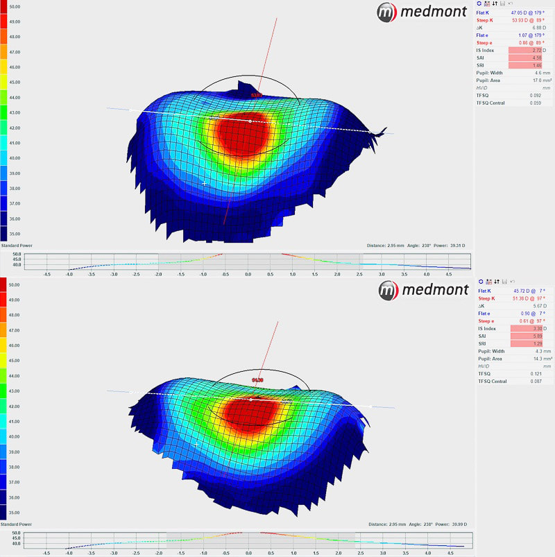 The distorted eye surface of this keratoconus case is clearly evident with 3D corneal topographical imaging. EYECARE CONCEPTS — ADVANCED EYE CARE & CONTACT LENS PRACTITIONER — MELBOURNE
A young lady in her 20s came to see us recently after having a sore red eye and blurred vision for about 5 days. She had been using the common over-the-counter antibiotic eye drop Chlorsig, typically used for a mild case of bacterial conjunctivitis (pink eye). This was the appearance of her eye on presentation: 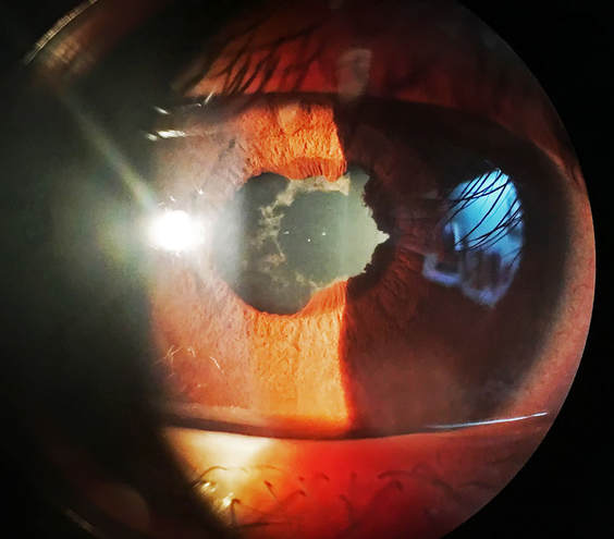 This young lady did not have conjunctivitis; she had a classic case of an eye condition called anterior uveitis, or iritis. It is an inflammatory condition of her iris, the coloured part of the eye that controls the pupil. It is a relatively uncommon condition that can affect a person of any age, typically a young to middle-age person. Symptoms usually include increased sensitivity to light (photophobia), eye pain or ache, redness in the eye, blurred vision and one pupil appearing smaller than the other.
Optometrists can diagnose anterior uveitis, and differentiate it from other kinds of pink eyes, by detailed examination of the eye with our slit lamp microscope. The necessary treatment for this condition is immediate and rapid control of the inflammation with a intense course of steroid eye drops (prednisolone acetate — Pred Forte) — typically every 15 minutes in the first 6 hours, then hourly dosing while awake, and reviewed the following day. A strong pupil dilation eye drop (eg. atropine) is also used to keep the pupil enlarged to prevent the inflammed iris from sticking to the front of the eye's natural lens (posterior synechiae). In this case as the iris had already adhered to the lens she had some permanent damage to her iris and pigment spots left on her lens. If this kind of eye inflammation is left untreated or not properly managed, vision loss and glaucoma (high eye pressures causing optic nerve damage) can result. With our treatment of strong steroid eye drops and pupil dilation drops she had relief from her eye pain within a short period of time. And at her next-day follow up the inflammation and redness had already subsided significantly with treatment. After continuing with treatment and allowing the inflammation to completely resolve, we referred her to see her GP for blood tests as anterior uveitis is a eye condition that has a strong association with other systemic inflammatory or autoimmune disorders. The take-home message from this case is that if you have a concern about your eye, please see an eye care professional who can make a sound clinical diagnosis and give you the treatment you need right away. Not all pink eyes are common conjunctivitis — if you have a painful (more than just an irritation) red or pink eye, light sensitivity or blurred vision you should see either a therapeutically-endorsed optometrist (an optometrist who is trained and qualified to prescribe medicines) or an ophthalmologist (eye specialist) without delay. And if you've been using an antibiotic eye drop such as Chlorsig for a pink eye but it doesn't show improvement, please seek further opinion for your eye promptly. For an after hours emergency or if you are unable to find a suitable eye care practitioner, the Royal Victorian Eye & Ear Hospital in East Melbourne would be the place to go. EYECARE CONCEPTS — THERAPEUTIC OPTOMETRIST — KEW EAST, MELBOURNE A pleasant young man was recommended by his mother to come for an eye check up with us recently. He had been seen within the last 3 years by another optometrist and has only a mild distance vision problem that he uses glasses for. The reason for his eye test was his concerns about some lid bumps and styes that he had seen his GP about several months ago but had still not been resolved with antibiotics that he was prescribed with. He had not noticed any vision changes and was still happy with his current glasses for driving. A comprehensive eye examination was carried out in our clinic, checking his vision, glasses prescription, thoroughly examining the front of his eyes in regards to the lid lumps and also a general eye health check. What was an entirely unexpected finding — and completely unrelated to his presenting issue — was his high eye pressures and potential changes to his optic nerves, indicative of early glaucoma. A visual field test — examining his peripheral (side) vision, which is implicated in glaucoma — thankfully did not detect any vision loss. He was promptly referred to a local ophthalmologist (eye specialist) who confirmed the glaucoma diagnosis and was treated with pressure-lowering glaucoma eye drops. By reducing his eye pressures to a lower and safer level, the risk of further long-term optic nerve damage and associated loss of peripheral vision is reduced. We will continue to monitor his eye health, eye pressures and vision for changes. What this case highlights is the importance of regular comprehensive eye tests for all. Everyone should be tested at least every 2 years regardless of whether one feels there is any change in eyesight or wears glasses or not. People over the age of 60, or has a family history of eye disease or additional general health risk factors such as diabetes, should be tested annually. Many eye conditions can develop without necessarily causing vision symptoms. For this young man — only in his early 40s (much younger than a typical glaucoma patient) — it was fortunate, in a sense, that because of his eyelid bumps that he came to see us for an eye examination. Otherwise he may not have had another eye check up for some time, and his high eye pressures could have led to some losses in his peripheral vision by that time. Eye care is much more than about glasses. If you haven't had your eyes checked for a while, book a comprehensive eye examination to ensure your eyes are healthy. And if you have any eye related concerns, see your friendly local optometrist right away — while GPs are fantastic at what they do for your general health, an optometrist is better placed to look at your eyes with our advanced eye diagnostic equipment. Comprehensive eye health checks here at Eyecare Concepts are bulk billed, so there are no out of pocket costs to you. Early detection is key to preventing vision loss. 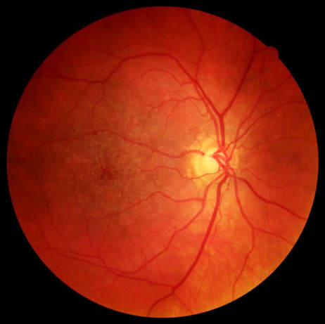 Glaucoma causes optic nerve damage, typically from high eye pressures. Case study published with patient's consent.
EYECARE CONCEPTS YOUR LOCAL EYE HEALTH EXPERT — KEW EAST, MELBOURNE |
AuthorPhilip Cheng - B.Optom (Melb) Ocular Therapeutics (GCOT). Optometrist at Eyecare Concepts Kew East, Melbourne. An experienced eye care & contact lens practitioner with expertise in myopia control & orthokeratology. Archives
August 2018
Categories
All
|
 RSS Feed